| Slide 52:
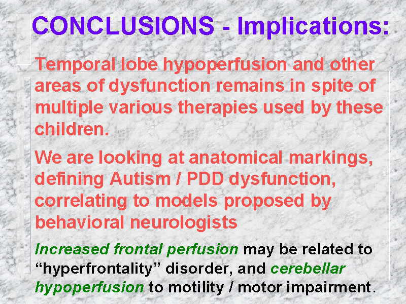
So the conclusions from this were: temporal lobe hypoperfusion and other
areas of dysfunction remained in these children in spite of multiple, various therapies
being used on these children. Being in southern California, I have the benefit or
non-benefit, whatever you want to call it, of children being treated with multiple
metabolic remedies. Certainly it’s Lovaas territory, ABA. At UC-Irvine, IVGG by Dr.
Gupta. What was striking was that any of these children that came to me, had done those
therapies – I would run a NeuroSPECT scan and these children still had temporal lobe
hypoperfusion.
We are looking at anatomical markings that define Autism/PDD as a model
that is consistent with behavioral neurologists. Now, what does that mean? I can’t do
this but when Dr. Miller looks at your children’s scans, he can define what is
working and what is not working. He can tell you what your kids are like. He can tell me
whether they are functioning right brain/left brain. We are looking at models that make
total sense.
One of the keys, and some of you have heard this before, Dr. Mena was
reading three primary areas there in the report I presented; decreased bloodflow in the
temporal lobe, decreased bloodflow in a touch of the parietal / occipital area, and
decreased bloodflow in a touch of the cerebellum. Dr. Mena, as a nuclear radiologist, was
reading these findings by the numbers. When I met Dr. Miller, he took 36 scans and
shuffled them like a deck of cards into mild, moderate, severe. He jokingly said to me
that he doesn’t read the numbers, he looks at the scans. Dr. Miller was able to bring
together that these areas that Dr. Mena was reading were interconnected anatomical tracks.
That’s something Dr. Mena had no way of knowing. That was the end of my ever worrying
about our spect scan data. Between that and Dr. Muntz’s report, it makes it easy to
tell you that we are looking at your children’s brains.
The increased frontal profusion may tie into hyperactivity, or as Dr. Mena calls it,
hyperfrontality disorder. The cerebellar hypoperfusion corresponds to problems in motility
or motor impairment. |